
Ankle xrays Don't the Bubbles
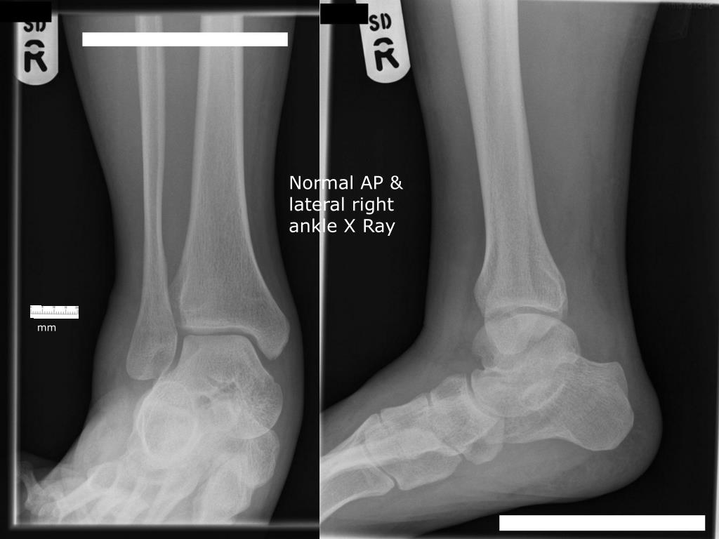
A standard ankle x-ray series consists of the AP, lateral and a 15 degree internal oblique (aka Mortise View) [2]. Figure 1: Example of a normal ankle series. Case courtesy of Andrew Murphy, Radiopaedia.org

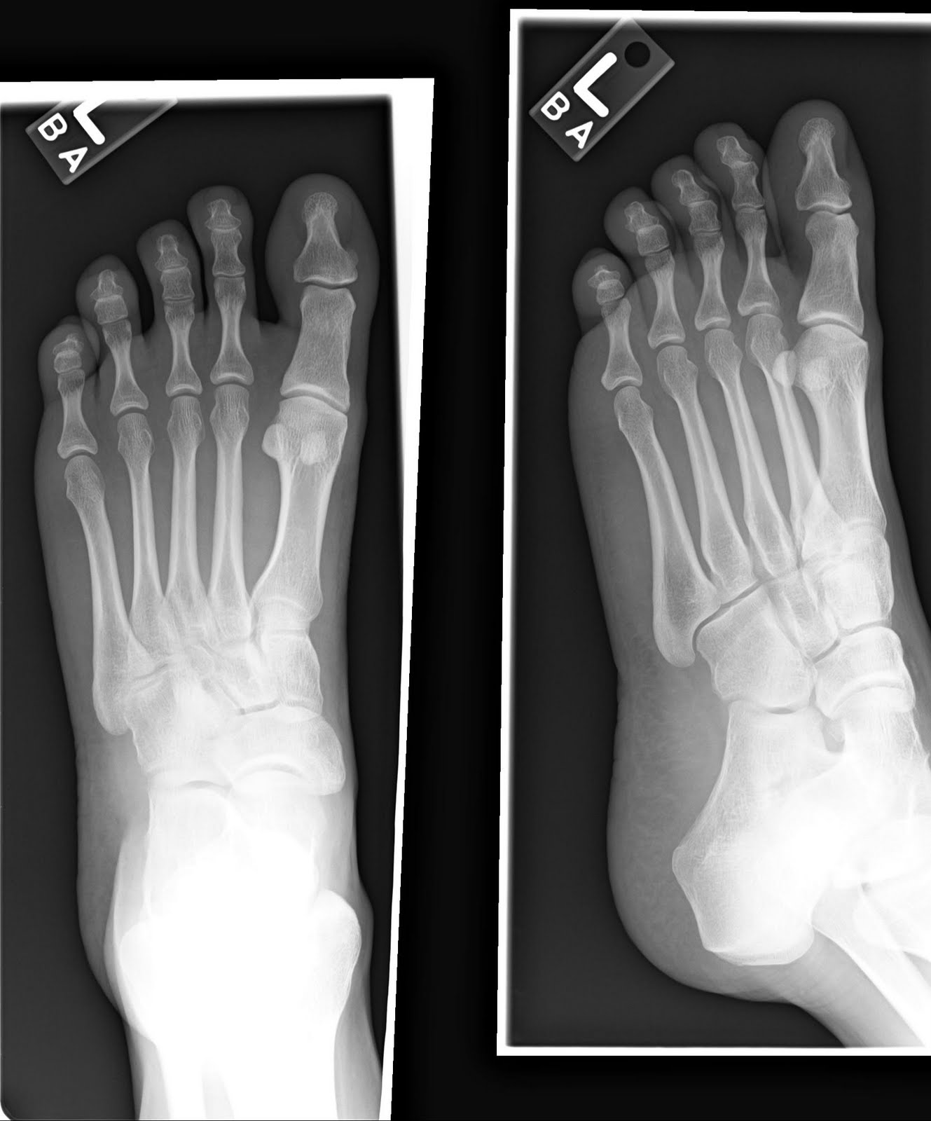
Normal Foot X Ray Normal foot series Image Check you have the right
There are three main sets of ligaments: Medial: deltoid ligament Lateral: posterior talofibular, anterior talofibular and calcaneofibular ligaments Syndesmotic ligament From Radiology Masterclass Ankle views An x-ray of the ankle will have three views - AP, mortise, and lateral.

Image
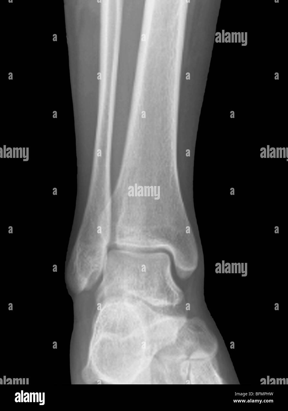
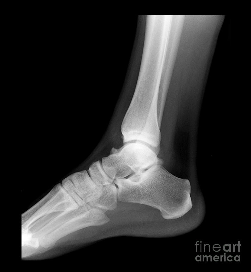
Normal ankle x-rays Case contributed by Ian Bickle Diagnosis not applicable Share Add to Citation, DOI, disclosures and case data Presentation Twisted ankle. Too much pop consumed. Patient Data Age: 35 years Gender: Male x-ray Frontal Lateral Normal appearances. x-ray Collated image of above. Normal examination. Case Discussion

Ankle X Ray Anatomy
X-ray technology is used to examine many parts of the body. Bones and teeth. Fractures and infections. In most cases, fractures and infections in bones and teeth show up clearly on X-rays. Arthritis. X-rays of your joints can reveal evidence of arthritis. X-rays taken over the years can help your doctor determine if your arthritis is worsening.
Normal ankle joint, Xray Stock Photo, Royalty Free Image 26886997 Alamy
Routine Radiographs These include a series of ankle and foot X-rays. ♦ Ankle series X-rays • Anteroposterior (AP) ( Fig. 2.1A) Fig. 2.1 (A and B) (A) Anteroposterior (AP) and (B) Lateral (LAT) views of ankle. • Lateral (LAT) ( Fig. 2.1B)
NORMAL FOOT 5
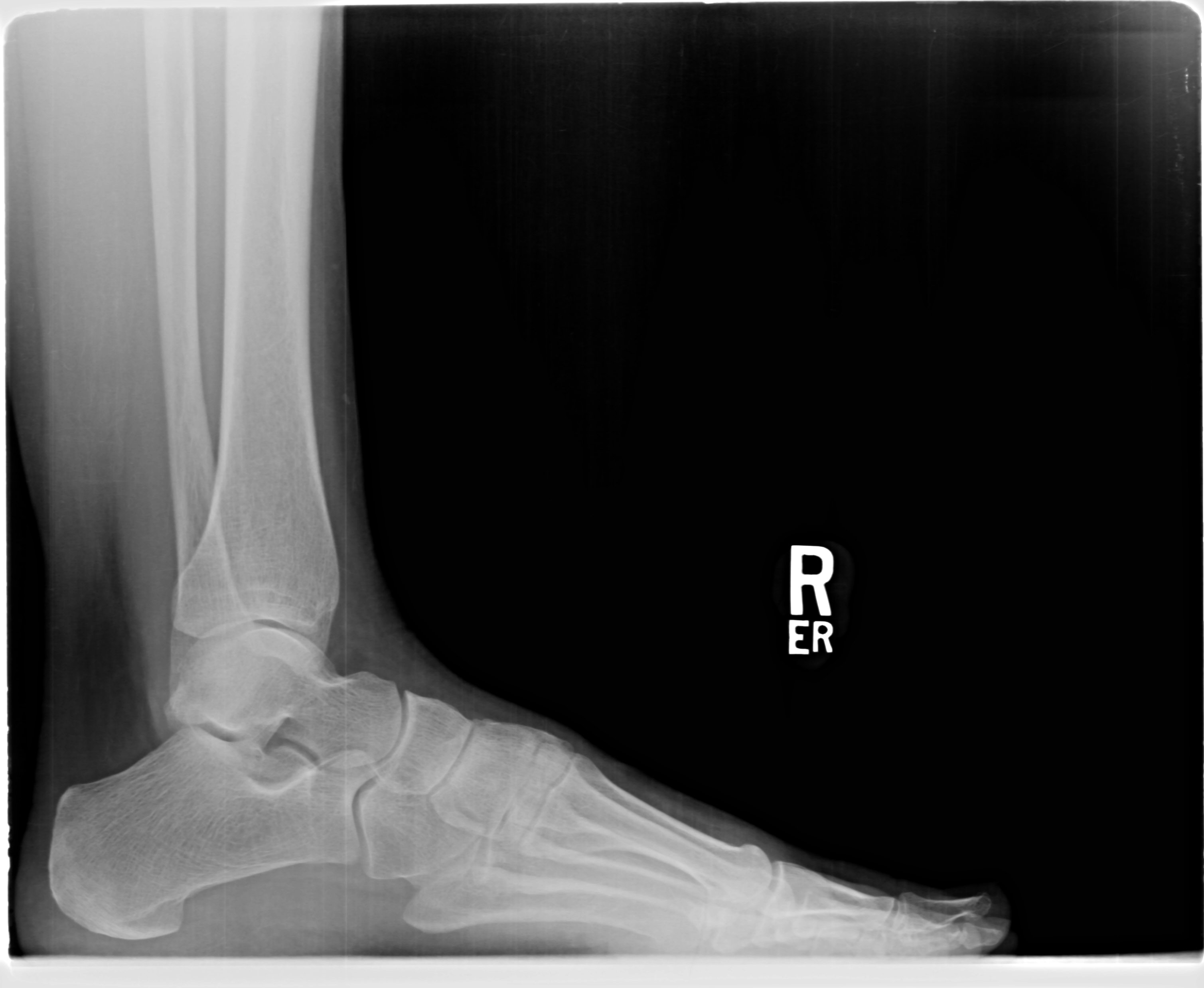
Correct side (right vs. left) Views In the United Kingdom, two views of the ankle joint are routinely performed: Mortise view: this is a modified anteroposterior (AP) view of the ankle in 10-20° internal rotation so that the medial and lateral malleoli are in the same horizontal plane and joint visualisation is optimised Lateral view

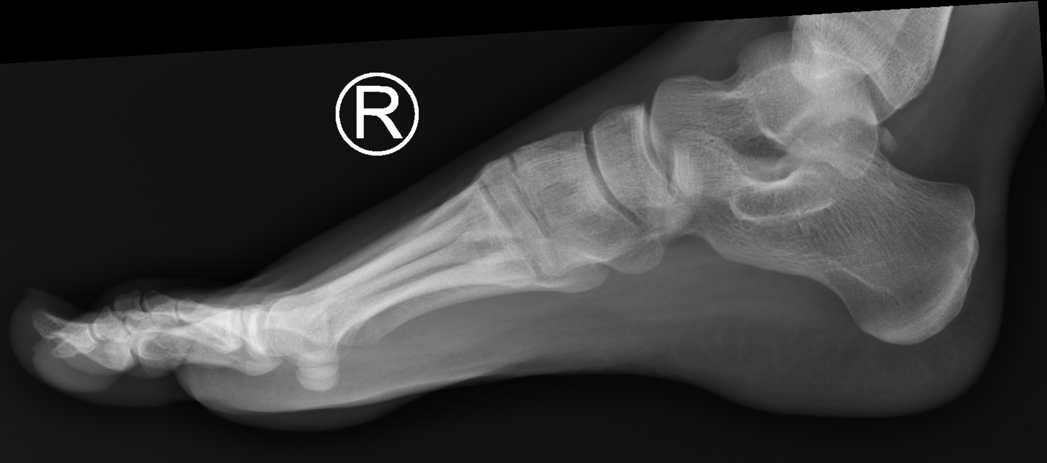
Normal ankle series Image
If questionable osseous findings noted on x-ray, consider CT to evaluate further. If x-rays are negative, consider MRI to search for occult osseous, ligament, or tendon injuries.. Note the normal fat density anterior to the ankle joint on the lateral view of the normal ankle ( Figure 11-1 C ).

RiT radiology When to Obtain Ankle Radiographs
Health Library / Diagnostics & Testing / Foot X-Ray Foot X-Ray A foot X-ray is a test that produces an image of the anatomy of your foot. Your healthcare provider may use foot X-rays to diagnose and treat health conditions in your foot or feet. Foot X-rays are quick, easy and painless procedures.

EMRad Radiologic Approach to the Traumatic Ankle
The true anteroposterior view of the ankle is often performed in the setting of ankle trauma and suspected ankle fractures in addition to the lateral and mortise views of the ankle. Other indications include: assessment of fragment position and implants in postoperative follow up evaluation of fracture healing
Assessing Heel Pain Diagnostic Ultrasound of the Foot and Ankle
same horizontal plane as the medial malleolus and both are parallel to the x-ray tabletop. The mortise view is the true AP projection of the ankle joint. Oblique projections, 1 plain radiograph. The radiographic appearance of the normal child's ankle is seen in Figure 21.11. The distal tibial epiphysis appears during the 2nd year of life.

normal right foot x ray Google Search Foot x ray Pinterest Foot pain
Introduction The ankle joint is one of the most commonly injured joints and the most common type of fracture to be treated by orthopedic surgeons. [1] The estimated incidence of ankle fractures is approximately 187 per 100,000 people per year. [2]
PPT XRay Rounds (Plain) Radiographic Evaluation of the Ankle PowerPoint Presentation ID
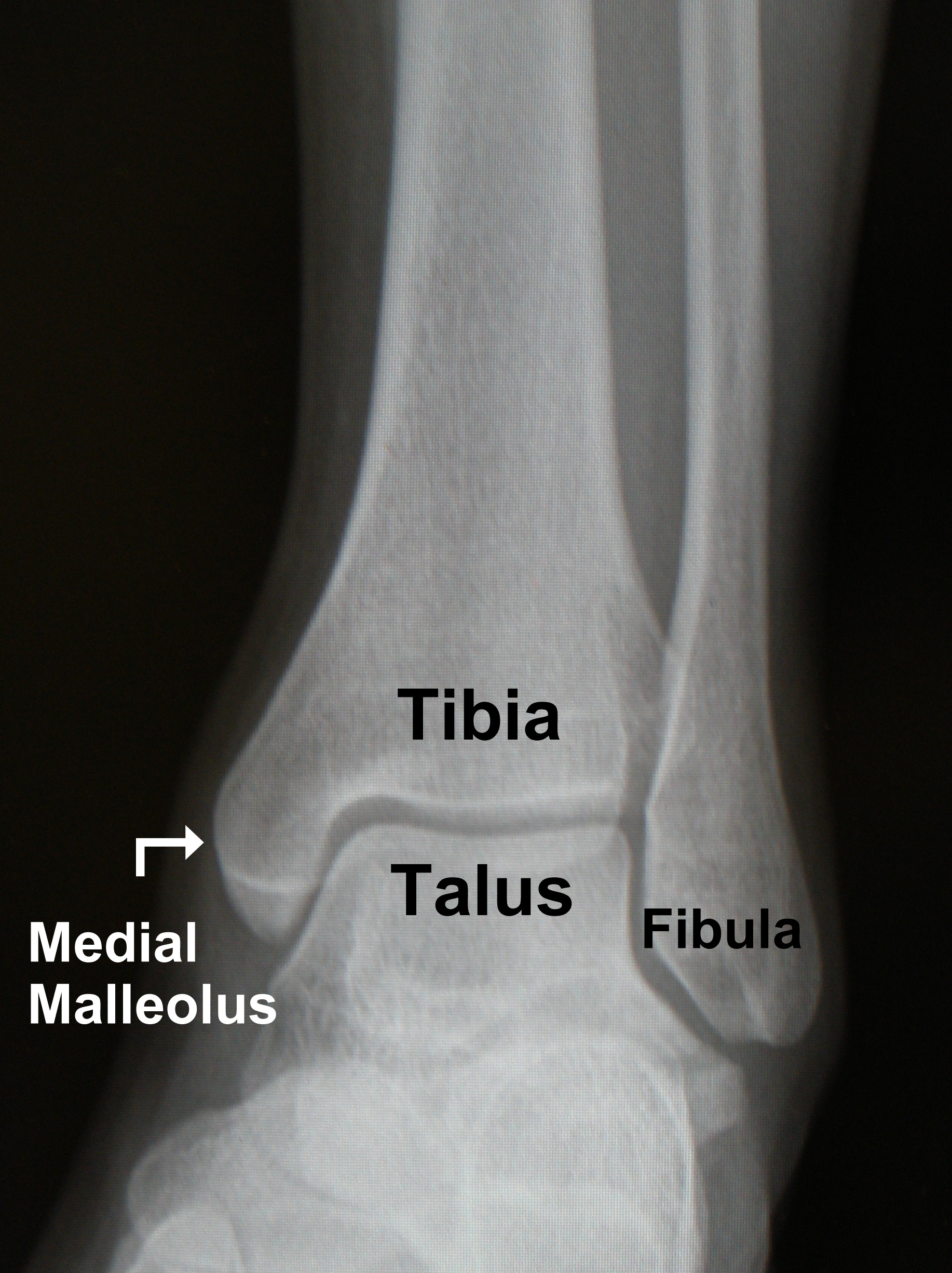
Ankle anatomy - Normal AP 'mortise' The weight-bearing portion is formed by the tibial plafond and the talar dome The joint extends into the 'lateral gutter' ( 1) and the 'medial gutter' ( 2) The joint is evenly spaced throughout Ankle anatomy - Normal Lateral Hover on/off image to show/hide findings
Ankle Xray, Normal Photograph by Living Art Enterprises
Indications This projection aids in evaluating fractures, dislocations and joint effusions surrounding the ankle joint, and helps to assess the severity of a calcaneal fracture by measuring the Böhler angle and Gissane angle. Patient position patient is in a lateral recumbent position on the table
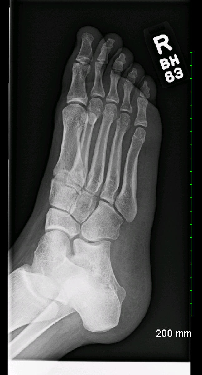
Normal Foot X Ray Normal foot series Image Check you have the right
Recognise normal variants and their significance (eg, accessory ossicles) Ottawa rules . These describe the requirements for plain x-rays within the clinical context of an ankle injury. They state that: an ankle radiograph is required only if there is pain in the "malleolar zone" and any of these findings:
Ankle Fracture FootEducation
Ankle Fracture Mechanism and Radiography. Robin Smithuis. Radiology Department of the Rijnland Hospital, Leiderdorp, the Netherlands. The ankle is the most frequently injured joint. Management decisions are based on the interpretation of the AP and lateral X-rays. In this article we will focus on:
NORMAL FOOT 7
Bony anatomy The ankle is a synovial joint composed of the distal tibia and fibula as they articulate with the talus. The distal tibia and fibula articulate with each other at the distal tibiofibular joint which is more commonly referred to as the tibiofibular syndesmosis (or simply the syndesmosis).